Transrectal Prostate ultrasound (TRUS)
Diagnostic value (10)
See Glossary of ultrasound Terminology
Ultrasound scanning of the patient with prostatitis is fundamental in the diagnostic and, as we shall see, therapeutic flow charts. Like everything else the scan must be done properly with the proper instrument. The patient’s bladder should be full (but not over-full) and the scan must be carried out by the transrectal route under basal conditions and during the dynamic phase of micturition, that is during urination. From experience we prefer to have the patient standing to facilitate micturition during scanning. For this reason the probe must be of the fine, new generation type so as to avoid bladder neck compression. As at least two planes are required for prostate scanning the probe must bi or multiplane and the crystal vibration in the range of 7-10 MHz. Only once these technical requisites are satisfied can we be sure of scanning the prostate properly.
 Under basal conditions the following ultrasound patterns must be monitored:
- prostate volume;
- capsule profile;
- tissue echogenicity;
- presence or absence of calcified fibrous formations,
- ejaculatory duct course and echogenicity;
- Cvein calibre in the peri-prostate plexus
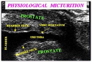
During micturition (See photo )the following patterns must be monitored:
- profile and echogenicity of the bladder floor (trigone);
- elasticity and morphology of the bladder neck;
- distension capacity of the prostatic urethra;
- profile and echogenicity of the prostatic urethra walls;
- profile and echogenicity of the veru montanum.